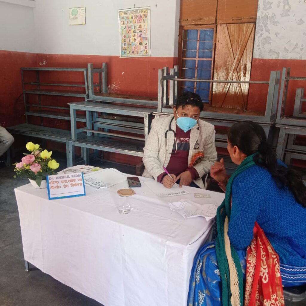
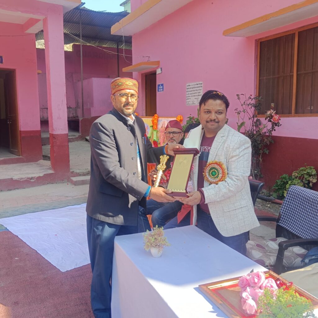
About Me
About Me

Professional background
Matriculation
-
- Institution: Nirmala Convent Senior Secondary School, Kathgodam, Nainital (U.K.)
- Board: CBSE
- Year of Passing: 2004
Intermediate
- Institution: Emmanuel Mission School, Kota, Rajasthan
- Board: CBSE
- Year of Passing: 2006

Post-Graduation (DNB Pulmonary Medicine/TB and Respiratory Diseases)
- Institution: Rajan Babu Institute of Pulmonary Medicine and Tuberculosis (RBIPMT Hospital), Delhi
- Board/University: Diplomate National Board (DNB)
- Year of Passing: 2018
Graduation
- Institution: Sri Guru Ram Rai Institute of Medical and Health Sciences (SGRRIM & HS), Dehradun, Uttarakhand
- University: H.N.B. Garhwal University
- Year of Passing: 2012
Professional background
Assistant Professor
GMC HALDWANI
- Nov 2019 – Nov 2022
- Teaching and clinical responsibilities at Government Medical College, Haldwani.
Senior Resident Doctor
RBIPMT Hospital, GTB Nagar, Delhi
- Clinical duties in a tertiary care setting, concentrating on pulmonary medicine and related fortes.
Medeor Hospital, Qutub, Delhi
- Clinical role in respiratory medicine and related fields
Junior Resident Doctor
RBIPMT (MCD run 1150 bed tertiary care hospital), Delhi
- 1 April 2015 – 31 March 2018
- Three-year residency concerning clinical training and patient care.
Rotatory Internship
Government District Hospital, Rampur (UP)
- 30 March 2012 – 29 March 2013
One-year comprehensive internship providing foundational medical experience
Education
DNB (Pulmonary Medicine)
- October 2018
Completed practical exams, specializing in pulmonary medicine
Skills
Dr Kedia possesses vast clinical skills in pulmonary medicine, including diagnosing diseases based on symptoms and clinical findings, managing cardiorespiratory emergencies, and prescribing medications in outpatient settings.
His procedural expertise encompasses various interventions such as pleural taps, tube insertions, fluid drainage, and bronchoscopies, with experience in bronchial sampling and thoracoscopic procedures. Dr Kedia exhibits progressive command in deciphering and analyzing diagnostic tests essential to pulmonary medicine, including X-rays, CT scans (HRCT and CECT), arterial blood gases (ABG), spirometry, and lung volume reports, contributing significantly to accurate patient assessment and treatment planning.
Dr Kedia's Approach Towards Patient Care
Dr. Kedia exemplifies a holistic approach to patient care, seamlessly integrating compassionate care with personalized treatment plans. As a highly skilled lung and pulmonary specialist, he is proficient in diagnosing diseases by carefully evaluating symptoms and clinical findings. Dr. Kedia’s adeptness lies in his ability to listen attentively to patients, ensuring that each individual’s unique circumstances are considered in the diagnostic process. His commitment to compassionate care fosters a supportive environment, while his tailored treatment strategies address the specific needs of each patient, ultimately enhancing outcomes and promoting overall well-being.
Rare Case Treatment
Have A look At some Rare Cases!
Case 1 - A case of a ticking time bomb in the human body
In late August, a 64-year-old male from Champawat, Uttarakhand, with a history of smoking, hypertension, and coronary artery disease presented with suspected left-sided pleural effusion, which was refuted upon review of the chest X-ray. His symptoms included chronic chest pain worsening over the past week and lower limb claudication. A 2D echocardiogram revealed elevated right heart pressures and a mass compressing the pulmonary artery, prompting a CT thorax that diagnosed a large fusiform thoracic aortic aneurysm with extensive mural thrombus.
Due to renal concerns, MR angiography was conducted, confirming the diagnosis. Immediate referral to Medanta Hospital for cardiac surgery was initiated, but due to financial constraints, the patient left against medical advice and tragically passed away within 72 hours of initial evaluation.
This case highlights the critical role of accurate diagnosis and timely intervention in managing complex cardiovascular conditions, underscoring the challenges posed by socioeconomic factors in accessing specialized care and affecting clinical outcomes.
Case - 2
An anonymous patient, a 74-year-old female, underwent a bronchoscopy performed by Dr. Anshul Kedia at Sanjivani Hospital, Haldwani, on January 16, 2024. The procedure utilized a Hugemed M58 flexible video scope with anaesthesia administered via 2% lignocaine solution, gel, and spray, along with sedation using 6mg diluted midazolam.
The examination revealed:
- Upper respiratory tract: Inflamed with a cobblestone appearance of the posterior pharyngeal wall, inflamed laryngopharynx, and epiglottis.
- Vocal cords: Inflamed but mobile with respiration.
- Trachea: Inflamed.
- Main carina: Widened.
- Right bronchial tree: Inflamed but patent.
- Left bronchial tree: Initially occluded with blood, cleared to reveal a large, friable mass in the left main bronchus.
Samples taken during the procedure included a bronchial wash from the left main bronchus for malignant cytology and an endobronchial biopsy from the mass for histopathological examination (HPE). These findings indicate a significant pathological condition in the left main bronchus, warranting further investigation and management.
Case - 3
An anonymous patient underwent a bronchoscopy on November 29, 2023, performed by Dr. Anshul Kedia. The procedure was conducted using Mr. Chanchal underwent a bronchoscopy on November 29, 2023, performed by Dr. Anshul Kedia. The procedure was conducted using a Hugemed M58 flexible video scope with anaesthesia administered via 2% lignocaine solution, gel, and spray, along with sedation using 4mg diluted midazolam. The examination revealed:
- Upper respiratory tract: Inflamed laryngopharynx and epiglottis.
- Vocal cords: Inflamed but mobile with respiration.
- Trachea: Inflamed.
- Main carina: Widened.
- Right bronchial tree: Severely narrowed with a lobulated, friable, silver-appearing endobronchial mass observed in the right main bronchus.
- Left bronchial tree: Inflamed but patent.